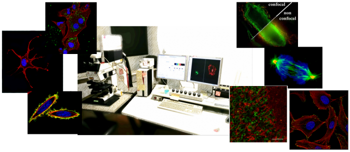
Microscopy Area
Confocal Microscopy
Confocal Laser Scanning Microscopy (CLSM) is a standard technology with the following advantages over conventional widefield microscopy:
- Removal of light from out-of-focus planes;
- Improved lateral (XY) and axial (Z) resolution;
- Ability to perform a series of optical sections from specimens;
The confocal microscope is a valuable tool for research because the system is able to obtain images with clarity and reasonable resolution, thus enabling 3-D reconstruction of the specimen from optical sections or series of images obtained.
The service involves the use of a latest generation Zeiss LSM980 confocal microscope with Airyscan 2 for high resolution imaging and a Leica TCS SP2 confocal microscope, both equipped with 5 laser sources at different wavelengths.
The Unit also provides equipment for sample preparation and maintenance prior to imaging, as well as resources for the data processing.
The 3D-imaging properties of the CLSM make it an ideal tool to study cell biology and life sciences.
|
Key applications areas include:
| ||
In addition, the Unit also provides basic protocols and all reagents needed to perform manually routine histological stainings (e.g. H&E and immune staining).
Personnel of the Unit assist scientist throughout the full process of microscopy experiments: design projects, processing data and analyzing images, and interpreting and shaping final results.
To ask for the service:
- preliminary contact the Confocal Microscopy unit staff;
- fill out the online form 'Service Request' briefly illustrating the scientific problem, type and number of samples, etc.;
- plan a meeting with the unit staff to discuss methods, costs, timing etc.
Contacts:
Serena Cecchetti, ☎ +39 06 4990 2966
Francesca Spadaro, ☎ +39 06 4990 6092
Where we are: building 20, floor 2, room 6
